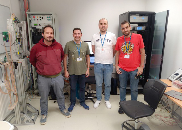
Source: CSIC Press Release
The Institute of Corpuscular Physics (CSIC-UV), the Institute of Structure of Matter (CSIC), and the Complutense University of Madrid (UCM) have developed the first entirely Spanish scanner for proton tomography.
A collaboration led by the Spanish National Research Council (CSIC) and consisting of the Institute of Corpuscular Physics (IFIC), a joint center of CSIC and the University of Valencia, the Institute of Structure of Matter (IEM-CSIC), and the Complutense University of Madrid (UCM), has successfully developed the first entirely Spanish scanner for proton tomography. This new device allows images to be obtained from the particles used in proton therapy, a new technique for treating cancer, thereby enabling better treatment dose planning. The initial results of this project were recently published in The European Physical Journal Plus.
Proton therapy is an advanced form of cancer treatment that uses protons. In recent years, it has gained popularity due to its advantages over conventional radiotherapy, as the characteristics of these particles, which make up the atomic nucleus, allow them to deposit almost all their energy in tumor cells while barely affecting healthy tissue. However, to properly plan the treatment, medical images of the patient are required.
Currently, these images are obtained with X-rays through what are known as computed axial tomographies (CT scans). However, the subsequent treatment is carried out with proton beams, not X-rays (composed of photons, the particles of light), which introduces uncertainties in treatment planning and dose calculation.
A possible solution to this problem would be to obtain images directly with protons. The innovative scanner developed by the Spanish collaboration led by CSIC is the first of its kind in Spain to achieve this goal, the researchers highlight. “Although it is currently a preclinical scanner that has obtained images of small mannequins, the results have been promising and have demonstrated the feasibility of the concept,” says Enrique Nácher, a CSIC scientist at IFIC who is leading this project.
The research team has combined a set of tracking detectors and a high-resolution energy scintillator to detect the residual energy of the protons. They used several mannequins irradiated with protons at a proton therapy center in Krakow, Poland. The mannequins were measured at different angles to obtain images reconstructed by filtered back projection, which were used to determine the scanner’s capabilities and validate its use as a proton computed tomography (proton-CT) scanner.
According to the results of the article, the scanner can produce medium-high quality images, with a resolution comparable to that of other state-of-the-art scanners. In the researchers’ opinion, if the system is properly scaled, it could be used to obtain images of patients before proton therapy, significantly improving treatment planning accuracy. “This would allow optimizing dose deposition in cancerous tissue while minimizing exposure to healthy tissue,” explains the IFIC researcher.
Resource Reuse
The proton scanner developed by the collaboration between the Institute of Corpuscular Physics, the Institute of Structure of Matter, and the Complutense University of Madrid was built by reusing instrumentation and materials from old prototypes of other nuclear physics projects that were no longer useful for their original purposes. This approach has maximized the reuse of resources without the need to invest in new instrumentation, promoting efficient and sustainable use of existing resources, its promoters highlight.


